Peri-implant diseases, including peri-implant mucositis and peri-implantitis, are increasingly observed across diverse US patient populations as dental implant use rises. This article synthesizes current epidemiology, diagnostic frameworks, and evidence-based management strategies to support clinicians in early detection, consistent diagnosis, and tailored treatment planning.
Introduction
Peri-implant diseases have emerged as a significant clinical concern in contemporary implant dentistry. With millions of dental implants placed in the United States and globally, clinicians must recognize the spectrum from reversible peri-implant mucositis to progressive peri-implantitis that jeopardizes implant longevity. This section establishes the clinical context, highlights key risk determinants, and frames the need for standardized diagnostic and management pathways.
1. Epidemiology of Peri-Implant Diseases in US Populations
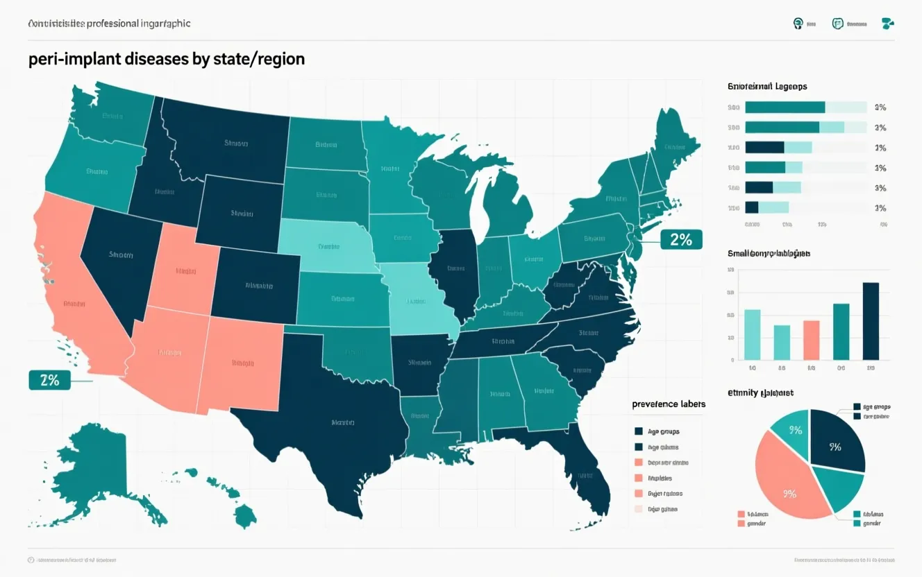
Definition and scope: Peri-implant mucositis is defined as an inflammatory lesion limited to the soft tissues surrounding an implant without concomitant loss of supporting bone, whereas peri-implantitis includes both soft-tissue inflammation and progressive loss of peri-implant bone. Prevalence estimates reported in the literature vary by study design, diagnostic criteria, and population sampled. Broad systematic reviews indicate peri-implant mucositis prevalence in implant patients often ranges from approximately 30% to over 60%, and peri-implantitis prevalence estimates commonly range from roughly 10% to 20% of implants — with some studies reporting higher rates depending on thresholds used for bone loss and clinical signs. For US-specific data, single-center cohorts, multi-center surveys, and registry analyses demonstrate similar variability; incidence is influenced by baseline periodontal status, maintenance frequency, and behavioral factors such as smoking.
Demographic and clinical variation: In US cohorts, age-related differences are observed but are typically modest when controlling for comorbidities. A history of periodontitis remains one of the strongest predictors of peri-implantitis across studies; patients previously treated for periodontitis show higher risk of future peri-implant bone loss. Smoking is consistently associated with increased prevalence and poorer treatment outcomes. Medical comorbidities, particularly poorly controlled diabetes mellitus, are linked to higher incidence and reduced healing capacity. Socioeconomic and access-to-care disparities in the US also influence maintenance adherence and therefore disease burden; urban versus rural differences can reflect access to periodontal specialty care and recall systems.
Incidence trends and longitudinal data: Longitudinal US registry and cohort data suggest that peri-implant disease incidence rises with time after implant placement, with a marked increase in risk after the first 3–5 years if maintenance is inconsistent. Key modifiable contributors to incidence include oral hygiene practices, prosthetic design that impedes plaque control, and inadequate peri-implant mucosal seal. Several US-based longitudinal studies (see PubMed indexation at PubMed) highlight that regular professional maintenance reduces incidence and severity; conversely, irregular or absent follow-up is a major driver of progressive disease.
2. Diagnostic Criteria and Classification Systems
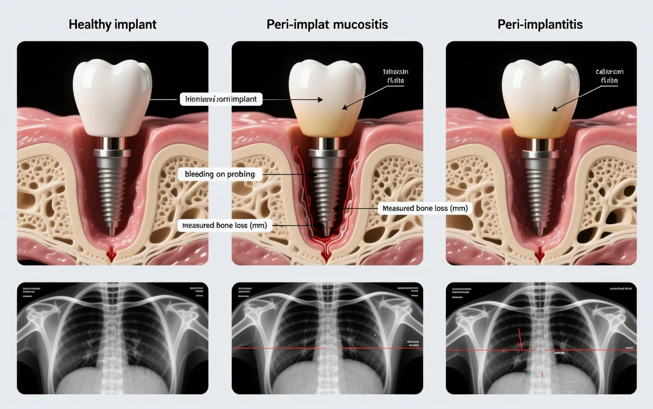
Core diagnostic parameters: Diagnosis of peri-implant diseases relies on a combination of clinical and radiographic findings. The primary clinical parameters include bleeding on probing (BOP) and/or suppuration, probing depth (PD), and changes in peri-implant mucosal appearance. Radiographically, vertical or circumferential bone loss relative to prior imaging or expected remodeling is critical for diagnosing peri-implantitis. However, the lack of universally agreed numeric thresholds (for example, how many millimeters of bone loss defines peri-implantitis) has produced variability across studies and clinical practice.
Comparison of major classification frameworks: Professional organizations and consensus groups — including the American Academy of Periodontology (AAP), the European Federation of Periodontology (EFP), and the European Association for Osseointegration (EAO) — have proposed criteria that share common elements but differ in specifics. The EFP/AAP consensus statements emphasize BOP as a hallmark of mucosal inflammation and recommend considering changes from baseline radiographs when available (AAP, EFP). The EAO provides clinical guidance on threshold values but acknowledges limitations related to probing variability and radiographic projection. In practice, sensitivity and specificity of these systems vary by clinician experience and the availability of baseline records; reliance solely on cross-sectional radiographs without baseline comparisons may under- or over-diagnose disease.
Standardization challenges and technological integration: Key challenges include variability in probing force and angulation around implants, differences in implant-abutment designs that affect soft-tissue position, and inconsistent radiographic techniques. Newer diagnostic adjuncts under investigation include cone-beam computed tomography (CBCT) for three-dimensional bone assessment, immunologic and microbial biomarkers (salivary and peri-implant sulcular fluid assays), and adjunctive quantitative digital probing devices. While CBCT can detect peri-implant bone defects more reliably than two-dimensional radiographs in certain contexts, radiation dose, cost, and artifact from metallic implants limit routine use. For practical clinical implementation, consistent recording of baseline clinical measures at implant placement and prosthesis delivery remains the most feasible method to improve diagnostic accuracy.
Practical recommendations for clinical consistency:
Obtain and archive standardized baseline periapical radiographs and clinical probing measurements at prosthesis delivery.
Use controlled probing force (approximately 0.25 N) and consistent probe angulation; document PD, BOP, and suppuration at each visit.
Consider CBCT selectively when two-dimensional imaging is insufficient or when surgical planning for regenerative therapy is contemplated (EAO, PubMed).
3. Non-Surgical Management and Adjunctive Therapies
Mechanical debridement: Non-surgical intervention is the first line for peri-implant mucositis and an initial step for peri-implantitis when defects are shallow and accessible. Mechanical debridement targets biofilm disruption and removal of surface contaminants. Instruments that have been evaluated include plastic curettes, titanium instruments, ultrasonic scalers with implant-compatible tips, and air-abrasive devices (air polishing). Clinical trials demonstrate that effective plaque control and thorough mechanical debridement often lead to resolution of mucositis; for peri-implantitis, non-surgical therapy alone achieves limited and variable bone-level improvement, particularly for defects with advanced bone loss.
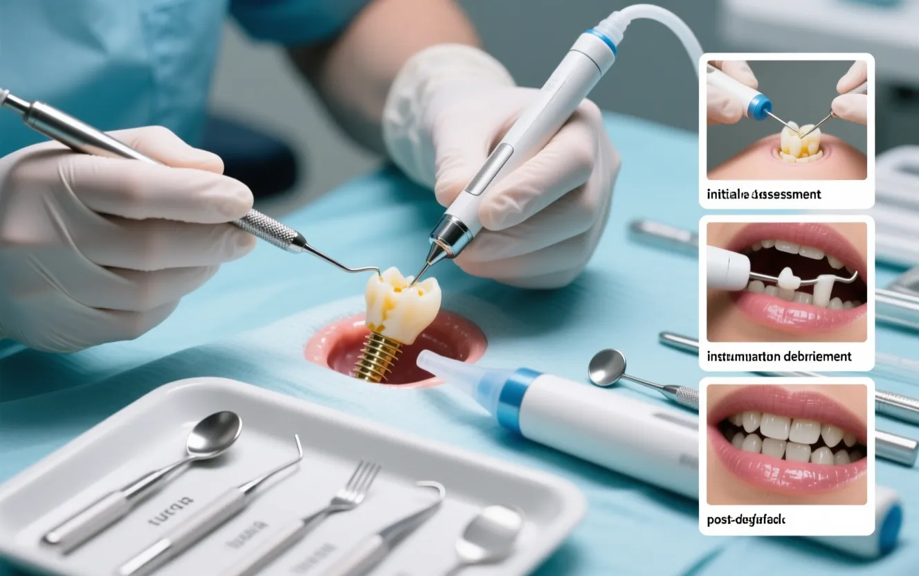
Instrument selection and efficacy: Titanium curettes or specially designed ultrasonic tips yield more effective removal of calculus and biofilm on implant surfaces compared to plastic curettes, which are less abrasive but may be inadequate for established deposits. Air-polishing with glycine or erythritol powders has demonstrated adjunctive benefits in reducing BOP and microbial load with minimal surface alteration. Clinicians should weigh the risk of altering implant surface topography against the need to decontaminate; in general, minimally abrasive methods that preserve surface integrity while reducing microbial burden are preferred for maintenance and early disease management.
Adjunctive antimicrobial therapies: Adjunctive antiseptics and antimicrobials frequently complement mechanical therapy. Chlorhexidine rinses (0.12%–0.20%) are commonly used as short-term adjuncts post-debridement. Local delivery of antimicrobial agents (minocycline microspheres, doxycycline gel) has shown additional reductions in pocket depth and BOP in some controlled trials, though effects may be transient and not consistently translate into radiographic bone regeneration. Systemic antibiotics may be indicated in selected cases with progressive disease, systemic involvement, or extensive defects, but routine use is discouraged due to resistance concerns and limited long-term benefit unless combined with effective surgical or mechanical decontamination.
Laser and photodynamic therapy: Laser-assisted therapies (e.g., Er:YAG, diode lasers) and photodynamic therapy have been investigated for implant surface decontamination. Results are mixed: some studies report improved clinical parameters when lasers are used adjunctively, while others demonstrate no significant advantage over conventional mechanical debridement. Parameter selection, operator experience, and heterogeneity of study designs contribute to variable outcomes; therefore, lasers should be considered adjunctive rather than primary treatments pending more robust evidence.
Clinical decision framework:
Peri-implant mucositis: emphasize patient-level plaque control education, professional mechanical debridement, and short-term antiseptic adjuncts as needed.
Early peri-implantitis with shallow defects: attempt combined mechanical debridement and adjunctive local antimicrobials; reassess response after 6–12 weeks.
Progressive peri-implantitis with deep or non-contained defects: refer for surgical evaluation, as non-surgical measures alone are unlikely to arrest bone loss.
4. Surgical Management: Regenerative vs Resective Approaches
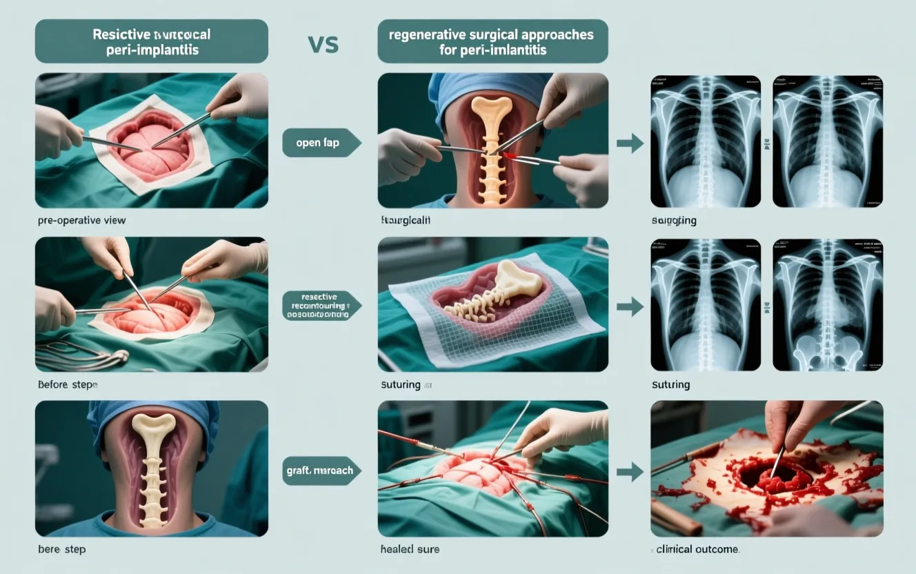
Indications for surgery: Surgical intervention is indicated when non-surgical therapy fails to resolve inflammation or when bone defects are deep, progressive, or compromise implant stability. The choice between a regenerative or resective strategy depends on defect morphology, esthetic demands, implant position, and patient factors.
Resective surgery and implant surface decontamination: Resective approaches include open flap debridement, apically positioned flaps, and recontouring of bone to create a maintainable architecture. The primary goal is to reduce pocket depth and eliminate inaccessible niches for biofilm. Surface decontamination methods used intraoperatively include mechanical (ultrasonic/brush debridement), chemical agents (chlorhexidine, citric acid), and adjunctive devices such as titanium brushes and lasers. Resective approaches provide predictable reductions in probing depth and BOP but may compromise peri-implant mucosal height and esthetics; survival rates are acceptable when proper maintenance is instituted, but long-term stability often requires strict plaque control and frequent professional recalls.
Regenerative procedures: Regenerative surgery aims to reconstruct lost peri-implant bone using graft materials (autograft, allograft, xenograft, synthetic biomaterials) and barrier membranes to facilitate guided bone regeneration. Indications favor contained defects (e.g., three-wall or circumferential defects) where scaffold and space maintenance are achievable. Published randomized and cohort studies report variable bone fill and clinical improvements; factors associated with improved outcomes include defect morphology, primary closure, thorough implant surface decontamination, and selection of appropriate graft/membrane combinations. Long-term comparative data suggest that regenerative procedures can achieve radiographic bone gain and improved clinical parameters in selected cases, but success is less predictable than regenerative therapy around natural teeth due to implant surface characteristics and ongoing microbial challenges.
Comparative outcomes and decision-making:
Resective surgery is often preferred for non-contained defects, shallow vestibular mucosa, or when esthetic compromise from regeneration is unacceptable.
Regeneration is preferred for contained defects in esthetic zones when bone reconstruction can support long-term soft-tissue stability.
Combined approaches (e.g., resective access followed by localized regeneration) may be appropriate for mixed defect morphologies.
Post-operative maintenance after surgery: Regardless of approach, post-operative maintenance is critical. Early and regular professional follow-up (e.g., 3-month intervals initially) combined with rigorous home plaque control predicts better long-term outcomes. Patient education on risk modification (smoking cessation, glycemic control) and prosthetic adjustments to facilitate hygiene are integral components of post-surgical care.
Conclusion
Synthesis of evidence: Peri-implant diseases are common in US patient populations and present a spectrum requiring accurate diagnosis and staged management. Epidemiologic data underscore the role of prior periodontal disease, smoking, systemic health, and maintenance adherence as primary risk determinants. Diagnostic inconsistency remains a major barrier to reliable prevalence estimates and timely intervention; obtaining baseline records and adopting consistent probing and radiographic protocols are pragmatic steps clinicians can take now.
Clinical recommendations: Prioritize prevention through patient education, prosthetic design that facilitates hygiene, and structured maintenance programs. Employ non-surgical debridement and adjunctive therapies for mucositis and early disease; recognize the limitations of these measures for advanced peri-implantitis and escalate to surgical consultation when indicated. Select resective or regenerative surgical strategies based on defect morphology, esthetic requirements, and patient-specific risk profiles, and ensure rigorous post-operative maintenance.
Future directions: The field requires standardized diagnostic thresholds adopted across professional organizations to harmonize research and clinical practice. Research priorities include validation of biomarkers for early detection, high-quality randomized trials comparing adjunctive therapies, and longitudinal US registry studies that stratify outcomes by demographic and socio-behavioral variables to inform equity-focused care. By integrating epidemiologic awareness, improved diagnostic consistency, and evidence-based treatment algorithms, US clinicians can better prevent implant complications and optimize long-term implant survival.